Transcrestal Sinus Floor Augmentation
with GUIDOR easy-graft
BY GUIDOR
Transcrestal Sinus Floor Augmentation
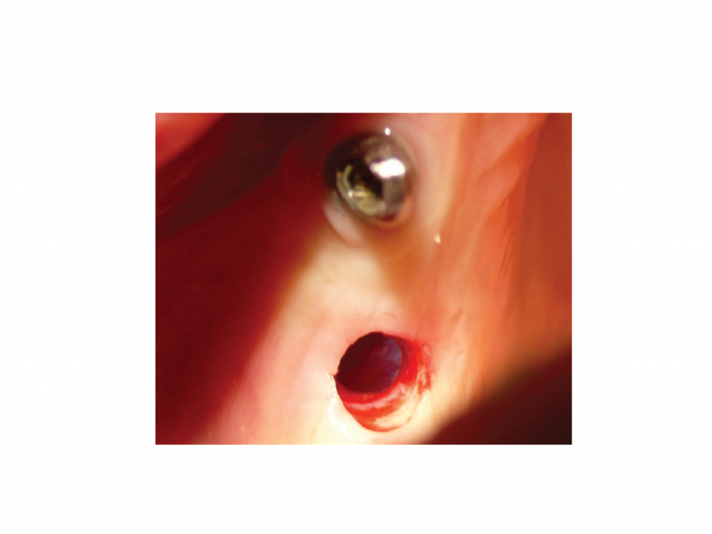
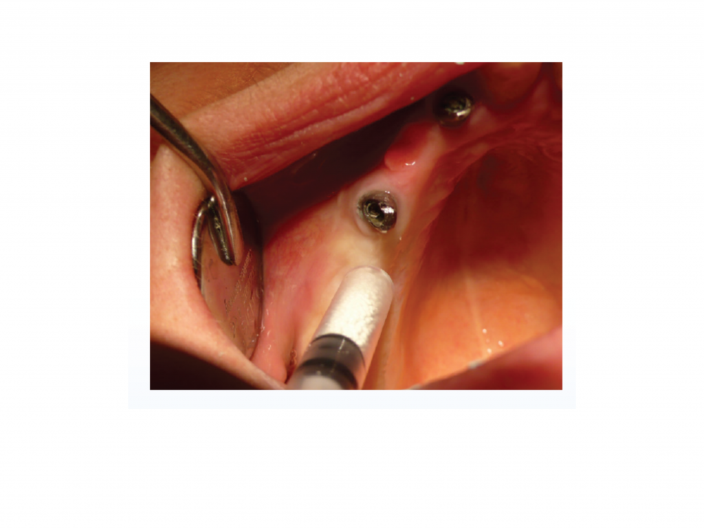
Fig. 3 Clinical situation with site opened and prepared for transcrestal sinus floor augmentation procedure
Fig. 2 Initial clinical situation
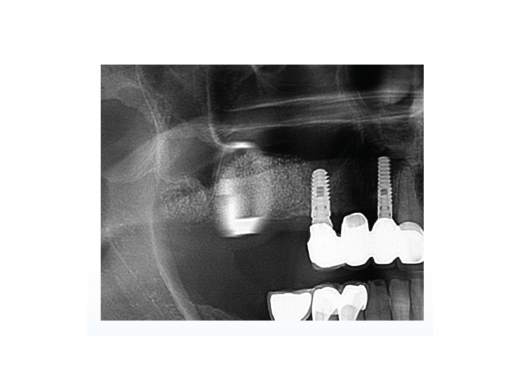
Fig. 1 Preoperative panoramic radiograph with residual bone height of 3 mm at position 16 (detailed view of situation)
Fig. 6 Detail of panoramic X-ray 6 months post op
Fig. 5 Detail of panoramic X-ray taken postoperatively after sinus floor augmentation
Fig. 4 Application of bone grafting material GUIDOR easy-graft CRYSTAL
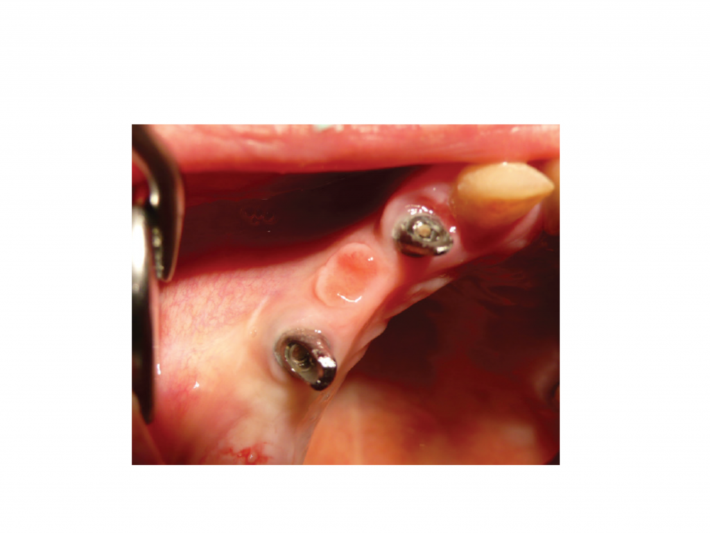
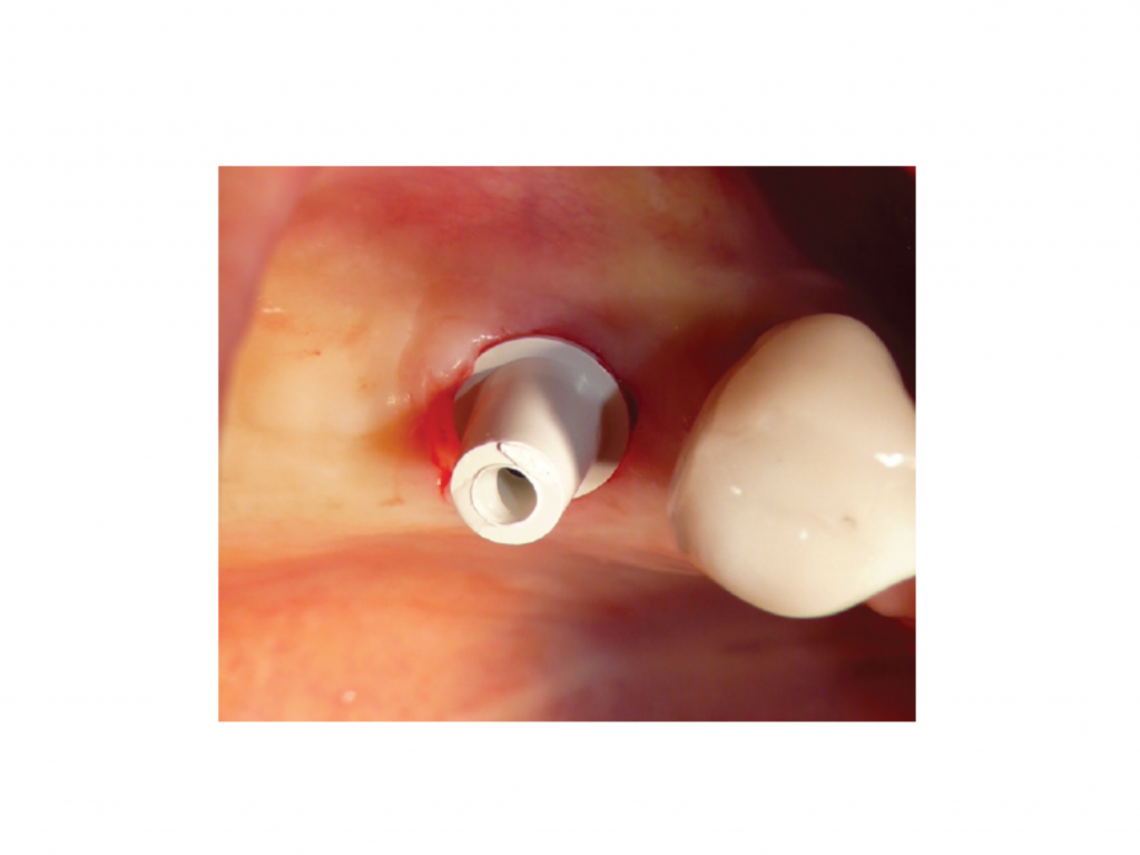
Fig. 9 Prosthetic treatment: clinical situation with installed PEEK abutment 4 months after implant placement
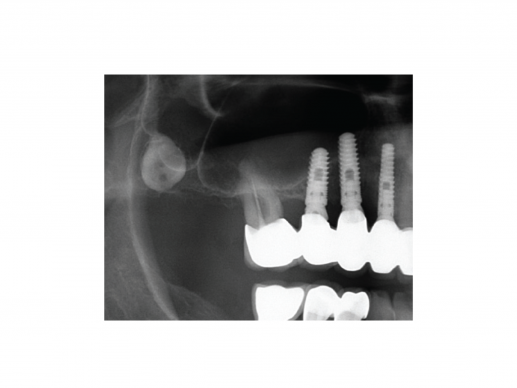
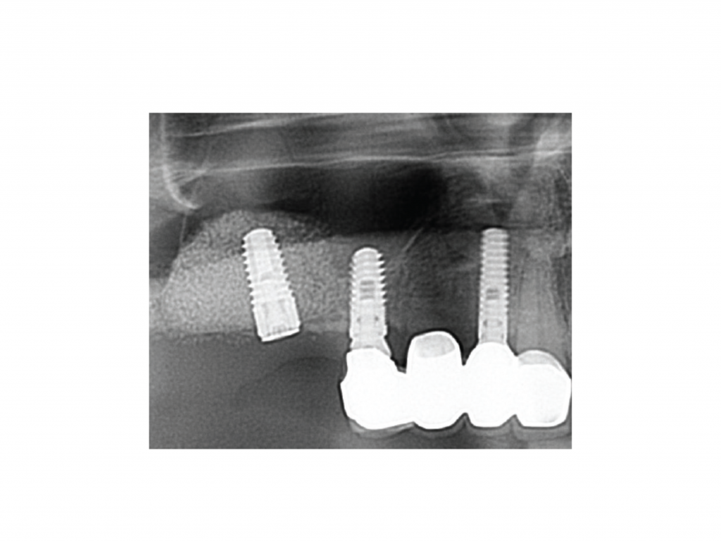
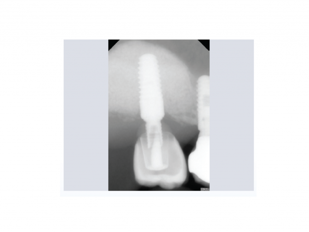
Fig. 8 Detailed view of panoramic X-ray after implant placement (Implant Blue SKY Classic)
Fig. 7 Implant placement 9 months
after sinus floor augmentation and histology showing GUIDOR easy-graft CRYSTAL with newly formed bone. (Histomorphometry: new bone: 27.7%, bone graft: 17.2%, connective tissue: 62.1%)
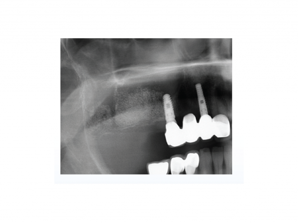
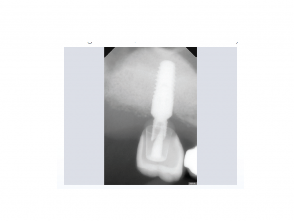
Fig. 12 Control X-ray, 2 years follow-up
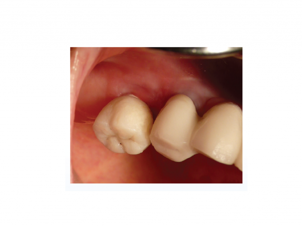
Fig. 11 Clinical situation after 2 years
Fig. 10 Radiographic view showing the final restoration in place