Tissue regeneration
with
PARASORB® RESODONT FORTE
BY
RESORBA

Tissue regeneration
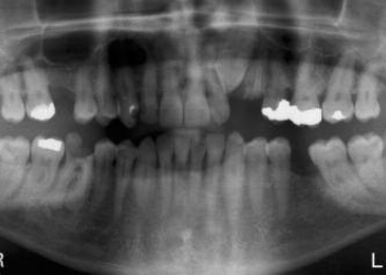
Presentation of three-dimensional bone defect from occlusal view
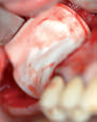
Presentation of three-dimensional bone defect from occlusal view
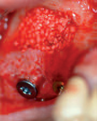
Inserted collagen membrane with perforation of the Schneiderian membrane in region 16 after insertion of implant
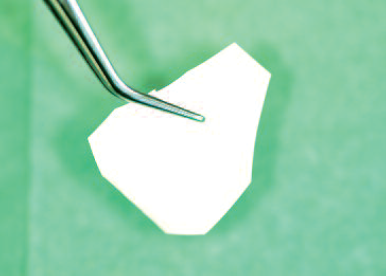
Collagen membrane cut to the defect size
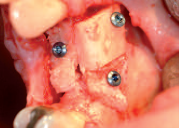
Bone
grafts fixed with micro-screws
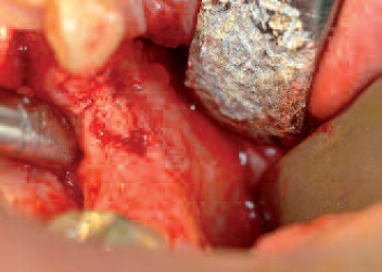
Presentation of three-dimensional bone defect from occlusal view
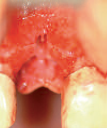
Presentation of bone defect (above all horizontally) after implant drilling in region 12
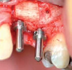
Inserted guide elements after removal of the osteosynthesis materials four months post operationem
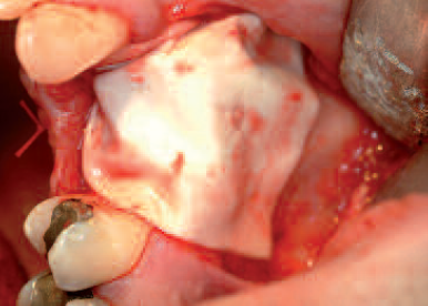
Augmented material fully covered by the collagen membrane
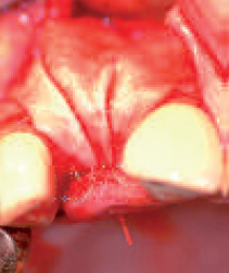
Collagen membrane positioned above the defect before wound closure
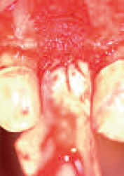
Autologous bone material positioned on top of the defect; collagen membrane palatally fixed with suture
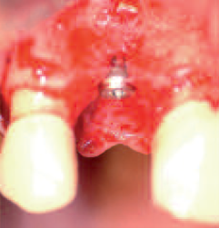
nserted implant with buccal bone defect at the level of the implant shoulder
